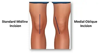
Medial Oblique Incision
2 months post op
Subvastus Total Knee Replacement
A knee replacement is a surgical procedure that restores the function of a damaged knee joint. More precisely, it is better described as knee resurfacing: the surgeon removes small amounts (millimeters) of worn bone and cartilage from the ends of the femur (thigh bone) and tibia (shin bone), and often the back of the patella (kneecap), then places metal and plastic components to recreate smooth, pain-free joint surfaces and proper alignment. The goal is to restore a smooth articulation so you can walk, climb stairs, and return to daily activities with less pain and improved function.
When it’s recommended: Knee replacement is considered only after nonsurgical treatments have failed to control pain and maintain function. Nonsurgical options typically include:
Activity modification and weight loss
Physical therapy and strengthening exercises
Anti-inflammatory medications (NSAIDs) and pain relievers
Corticosteroid injections into the knee
Viscosupplementation (hyaluronic acid) injections
Bracing and orthotics
Assistive devices (canes, walkers)
Who is a candidate: Patients with persistent, severe pain or disability from arthritis (usually osteoarthritis, sometimes inflammatory or post-traumatic arthritis) despite the above measures are candidates for knee replacement/resurfacing. Other considerations include deformity, instability, and impact on quality of life.
The operation and what to expect
Anesthesia: regional (spinal) or general anesthesia, often with nerve block for postoperative pain control.
Resurfacing: the surgeon removes a few millimeters of bone and cartilage and fits metal components to the femur and tibia, plus a plastic spacer to recreate the joint surface. The patella may be resurfaced selectively.
Duration: typically 35-45 minutes.
Recovery:
Most patients go home the same day (outpatient procedure). Overnight stay may be recommended for patients with significant medical problems, inadequate home support, or living alone without help.
Pain control uses multimodal methods: nerve block, oral medications, and ice.
Early motion: physical therapy begins the day of or after surgery. Walking with a walker or cane and range-of-motion exercises start immediately.
Typical timeline:
First 1–2 weeks: manage pain, control swelling, continue walking with assistive device as advised, begin home or outpatient physical therapy.
2 weeks: most patients walk with minimal or no assistive device and perform daily activities with less pain.
1-2 months: substantial improvement in pain and function; many return to low-impact activities.
3-4 months: full recovery of strength and endurance; final results of surgery are apparent.
Risks and longevity: All surgery carries risks: infection, blood clots, stiffness, nerve or vessel injury, ongoing pain, and need for revision surgery. Modern implants and techniques typically last 20–25 years or longer depending on activity level, body weight, and implant type.
Bottom line: Knee replacement is effectively a resurfacing procedure that removes small amounts of damaged bone and cartilage to create smooth joint surfaces. It is reserved for patients whose pain and disability persist despite nonsurgical treatments. Recovery is progressive, and most patients are safely treated as outpatients unless medical or social reasons require observation. Your surgeon will review individualized risks, expected outcomes, and the rehabilitation plan before proceeding.
Total knee replacement animation
A Different Approach to a Knee Replacement
The Subvastus Approach
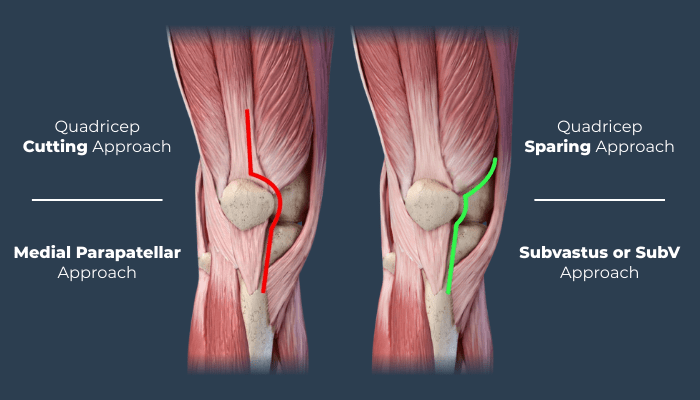
The Subvastus (also called the medial subvastus) exposure for total knee arthroplasty is a less invasive surgical approach that preserves the quadriceps tendon and fibers of the vastus medialis, in contrast to traditional medial parapatellar or mid-vastus approaches. This preservation leads to less disruption of the extensor mechanism, which can translate into earlier active knee motion, reduced pain with straightening and bending, and often a faster initial functional recovery.
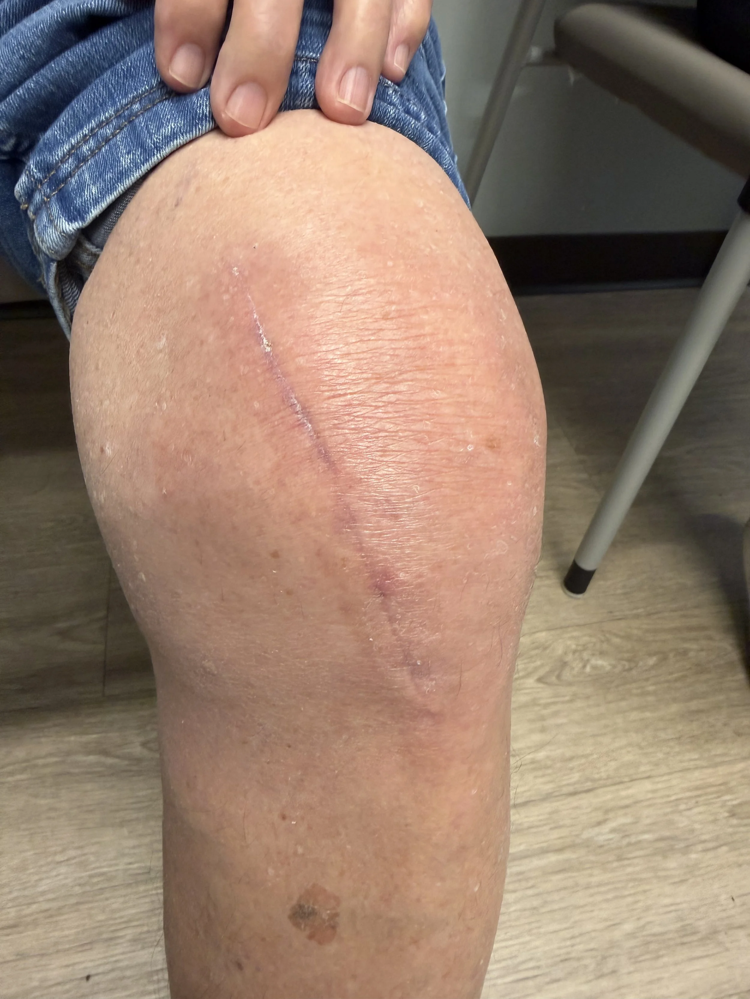
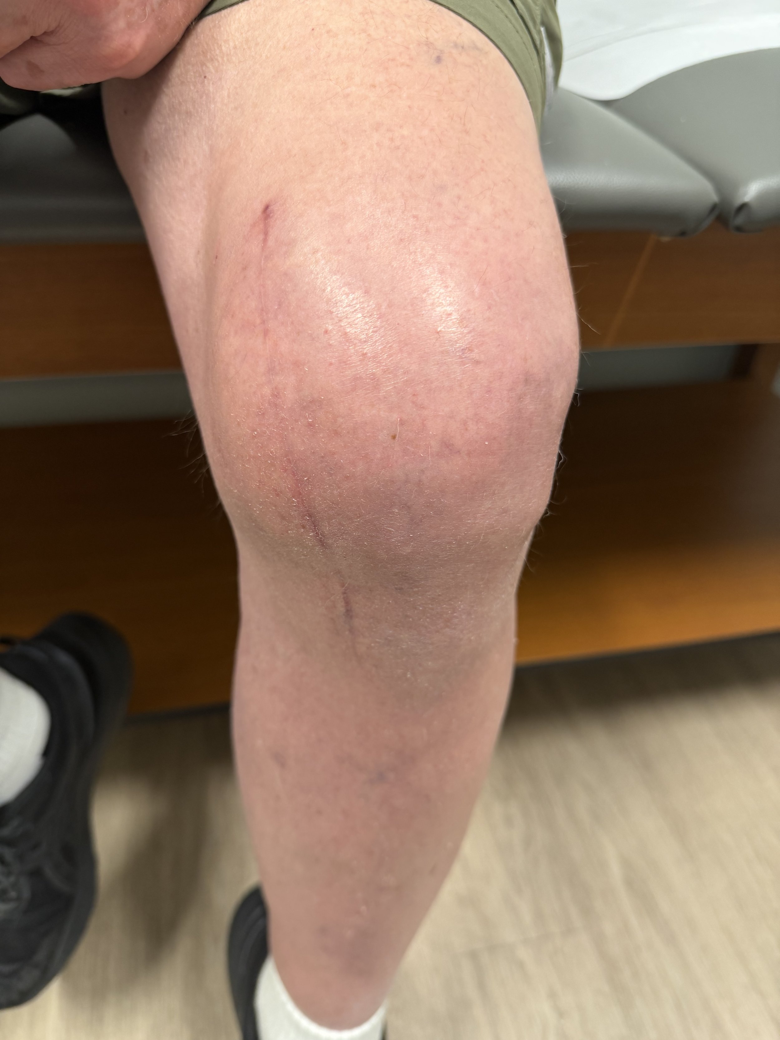
Medial Oblique Incision
2 months post op
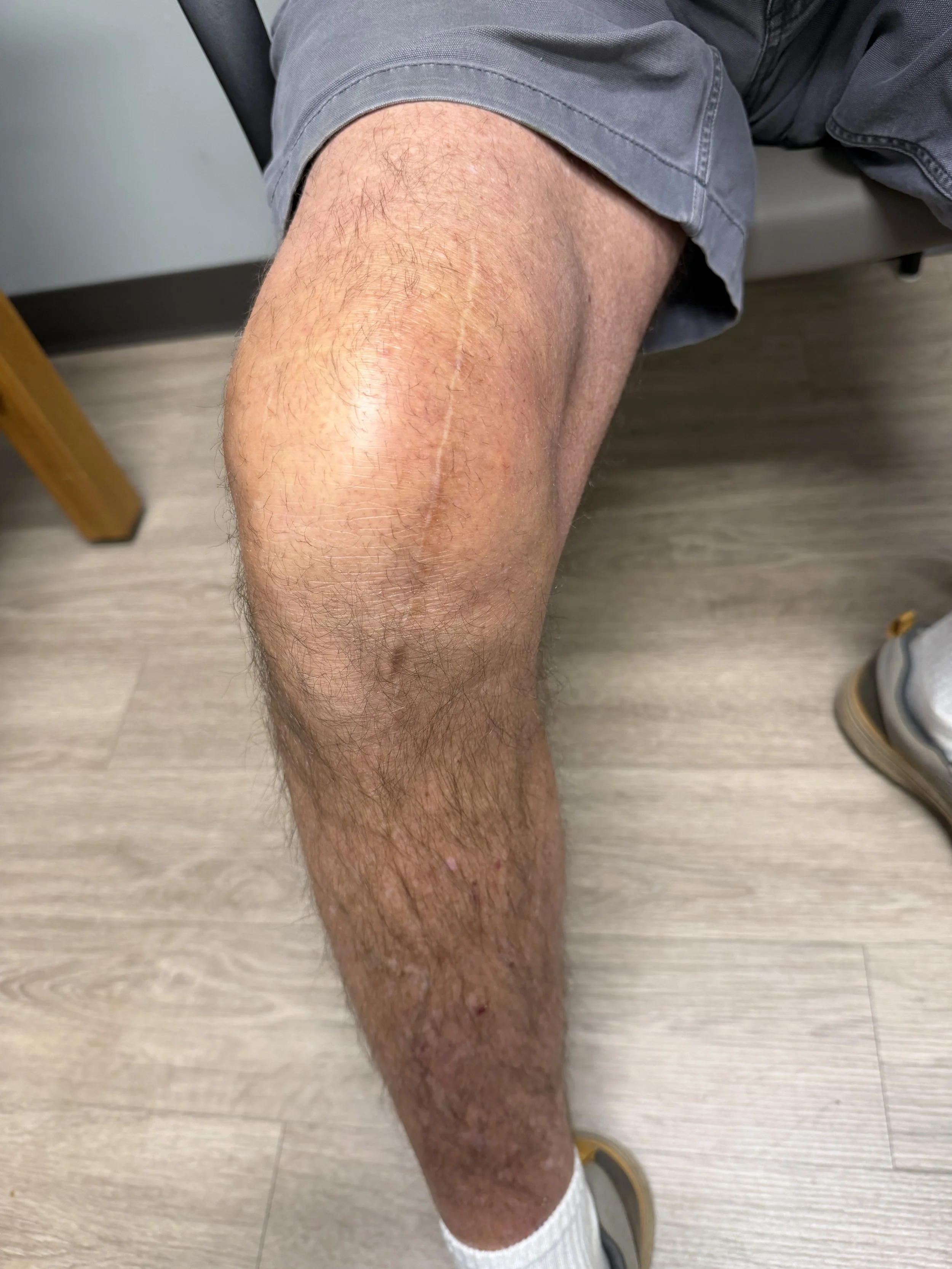
Medial Oblique Incision
12 months post op
Key differences in exposure and technique
Tissue plane: The subvastus approach develops a plane beneath (deep to) the vastus medialis muscle rather than incising the quadriceps tendon or splitting the vastus lateralis/medialis fibers. The muscle is gently elevated and retracted medially to expose the joint.
Extensor mechanism integrity: Because the quadriceps tendon and its continuity are maintained, the extensor mechanism is left intact. Traditional medial parapatellar approaches require detachment or incision through the quadriceps tendon or parapatellar retinaculum, and mid-vastus splits muscle fibers.
Patellar handling: The patella is everted much less (often subluxed laterally rather than fully everted) or can be left in place and shifted, reducing traction on the extensor mechanism and patellar blood supply.
Skin and soft-tissue incision: Skin incision is similar in length and location to the medial parapatellar approach, but deeper dissection follows a more medial and submuscular route.
Clinical advantages
Early range of motion (ROM): By sparing the quadriceps tendon and minimizing muscle disruption, patients frequently achieve active knee extension and flexion earlier in the postoperative period.
Less pain and improved function early: Reduced trauma to muscle and tendon correlates with lower early pain and better ability to perform quadriceps activation exercises, transfers, and ambulation.
Faster rehabilitation milestones: Patients often meet early goals (straight-leg raise, independent ambulation with assistive devices) sooner than with approaches that violate the extensor mechanism.
Potentially lower risk of extensor lag: Preserving the quadriceps continuity reduces the chance of postoperative weakness or extensor lag attributable to tendon/muscle injury.
*Individual results may vary
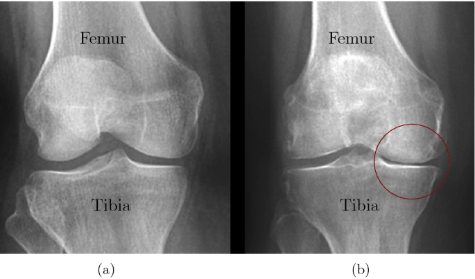
Arthritis is a process that involves the gradual loss of cartilage. To give you a simple analogy, think of it as wearing the tread off the tires on your car. If you put enough miles on your tires, the tires will wear all the tread off. One side of the tire may wear more than the other, just like one side of your knee may wear out before the other side. Patients will often ask “why is my knee hurting so much now if arthritis has been there for a long time?”. I’ll answer this using that same tire analogy. You have been wearing those tires down for a long time. Your mechanic might have even suggested some new tires because some were worn down completely, but you keep driving on them until one day you have a blow out. The process took some time, but eventually things fall apart.
A knee replacement would be better called a knee resurfacing. It is a surgical procedure that changes the surface on the end of the thigh bone (femur) and top of the shin bone (tibia). A few tools are used to remove the worn surface of the bone and then reshape the bone to fit the new metal implants. The implants are typically cemented to the bone and allow you to walk on it the same day. Most of our patients go home the same day as the procedure unless they have significant medical issues.
Phone: 706-839-4096